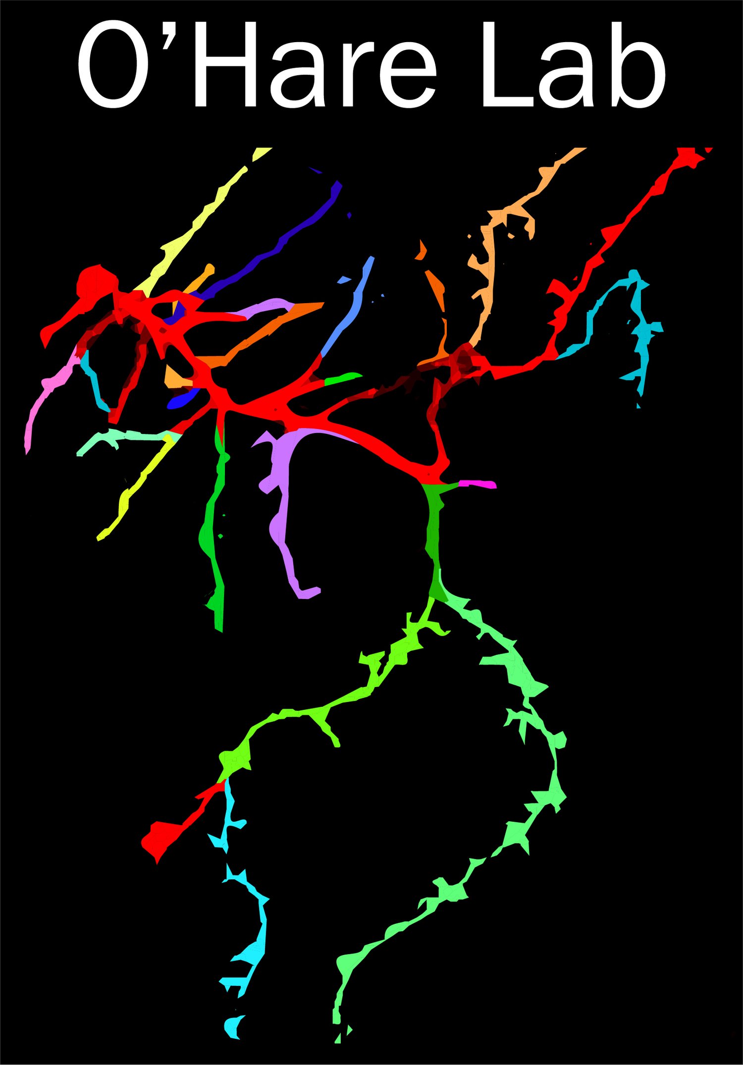
Our Mission
The O’Hare lab seeks to understand how individual neurons contribute to memory formation and storage. We pursue this goal by bridging the gap between two traditionally siloed fields of study: cellular/molecular neuroscience and systems neuroscience.

Cellular neuroscience focuses on the structure and function of individual brain cells (e.g., neurons) and has provided detailed insights into the molecular mechanisms of synaptic plasticity: the strengthening or weakening of connections between neurons that is thought to support learning and memory.
Systems neuroscience attempts to understand the brain as a machine of interacting systems (usually whole circuits) and has uncovered key computational principles for the formation and storage of new memories across many neurons.
These disciplines each require extensive sets of conceptual and technical expertise and, as such, an entire research lab typically falls within one discipline.
Why is bridging cellular and systems neuroscience disciplines essential to understanding learning and memory? Let’s consider our model system of choice: the pyramidal neuron from area CA1 of the mouse hippocampus:

This is a reasonably drawn cartoon of a pyramidal neuron from hippocampal area CA1. It integrates a continuous barrage of > 10k synaptic inputs into a binary train of action potentials that helps guide behavior. Impressive but not too complicated…right?

Now let’s consider that those inputs originate from different presynaptic circuits that transmit distinct, complementary streams of information - all of which are important. Things are getting complicated: the neuron’s action potential firing is a function of three distinct types of synaptic input. But that’s not all…

Each presynaptic circuit targets a different set of dendritic compartments that are biophysically distinct from one another! This nexus between systems- and subcellular-level organization is (1) critical to understanding how individual neurons learn to represent features of the environment, (2) largely uncharted in vivo, and (3) our core area of focus.

As a mouse explores a new environment, CA1 pyramidal neurons form new receptive fields that manifest as the neuron selectively firing when the mouse is in a particular location. These receptive fields are known as “place fields” and CA1 pyramidal neurons with place fields are known as “place cells”.
Place cells form rapidly and can just as quickly “remap” to new locations in a given environment or, when an animal moves to new surroundings, respond to a location in its new environment.
Place fields are considered a cellular substrate for learning and memory. Their name is deceptively simple - place fields conjunctively encode many features of an animal’s environment. Analogous receptive fields support episodic learning and memory in humans.
The vast amount of research into place fields falls mostly into two buckets: molecular mechanisms of synaptic plasticity in hippocampal pyramidal neurons (cellular neuroscience) and high-level analyses from in vivo imaging experiments monitoring the activity of many (100’s - 1,000’s) of neurons (systems neuroscience).
Herein lies the rub: despite the immense progress separately made with these two approaches, we still know very little about how new place fields form in the intact mammalian brain. By using place field formation as a model system to study how presynaptic circuits interact with specific dendritic compartments in individual neurons in vivo, we aim to uncover new general principles governing learning and memory.
Our Approach
Many of our experiments are designed to directly monitor and manipulate the activity of CA1 pyramidal neuron dendrites.
These experiments require maximally sparse labeling: the CA1 pyramidal layer is densely packed such that, even with 2-3 labeled neurons, it can be nearly impossible to correctly attribute a given dendrite to its parent cell body. In part for this reason, we often generate single-cell preparations using in vivo single-cell electroporation (SCE).
In short, we directly introduce DNA plasmids to individual CA1 pyramidal neurons in anesthetized mice and return days later to image those neurons at subcellular resolution in awake and behaving mice.

Diagram of SCE approach. Top: a long-tapered pipet is used to deliver plasmid DNA to an individual neuron. Bottom: 2-photon images before, during, and after SCE in the CA1 pyramidal layer.
Movie stepping down axially through an electroporated CA1 pyramidal neuron, starting at the soma and ending at the distal tuft dendrites.
A 3D reconstruction of the same electroporated CA1 pyramidal neuron.
Additional benefits of directly introducing DNA plasmids to individual neurons:
Flexibility: > 100k plasmids are readily available via Addgene and, when we can’t find what we need, we generate our own custom constructs via DNA subcloning.
Genetic manipulations: We can genetically manipulate individual neurons in adult mice in vivo using the Cre-loxP system and, in principle, CRISPR analogues suited to post-mitotic cells. Acute, single-cell manipulations circumvent developmental effects from constitutive manipulations (e.g. knockout mouse lines) as well as circuit-level effects from targeting many neurons at once (e.g. viral approaches).
Thus, SCE allows us to directly monitor and perturb an endless array of subcellular phenomena that the field has long thought to be essential for learning and memory but have proven experimentally inaccessible in the intact brain.
For recent examples of SCE-enabled subcellular interrogation of place field formation in mouse area CA1, see:
O’Hare, et al 2022 (Science). PubMed Central (open access)
Published protocol (Nature Protocols); PubMed (open access available Dec. 2025)

Additional techniques used in the lab
Virtual reality-based behavior

Volumetric multiphoton imaging and optogenetics

Population calcium imaging

Slice electrophysiology
Using the approaches previewed here and others, our lab aims to uncover how numerous subcellular processes contribute to learning and memory across anatomical and functional scales:

Initial Projects
-
A major initial focus of the lab is to uncover exactly how intracellular calcium release (ICR), from the endoplasmic reticulum, promotes place field formation in the dendrites of CA1 pyramidal neurons.
Dr. O’Hare’s postdoctoral work established the first causal link between ICR and the emergence of dendritic feature selectivity (of which place fields are an example).
Having established a critical role for ICR in place field formation, we will step back to address three fundamental questions using all-optical methods in vivo at subcellular resolution:
When does ICR occur in dendrites of CA1 pyramidal neurons?
Where does ICR fit into the chain of subcellular events culminating in the emergence of a new place field?
How is ICR engaged across pathway-specific dendritic compartments and its two canonical release mechanisms?
Each of these aims provides an exciting thesis or postdoc project with wide-ranging conceptual and technological training opportunities.
-
As explained in the sections above, we strive to understand how individual neurons integrate diverse synaptic inputs from multiple circuits to form new, information-rich receptive fields during learning.
Our research benefits from many recent technological advances, particularly related to in vivo imaging. At the same time, we actively develop new methods and approaches in the lab with the goal of improving our own capabilities as well as those of the rest of the field. Example methods-based projects include but are not limited to:
A comprehensive, easy-to-use, object-oriented Python package for managing, processing, analyzing, plotting, and disseminating in vivo imaging datasets - including those arising from multi-channel, volumetric dendritic imaging experiments. This package will interface with Neurodata Without Borders.
An automated whole-neuron reconstruction software suite that can trace, fill, and segment dendritic arbors based on noisy z-stack images such as those acquired in vivo at depth through light-scattering tissue. This software suite will be integrated into our analysis package described in (1) to better enable researchers to pair form with function.
We are developing new imaging approaches to investigate hippocampal function across subcellular- and circuit-level scales. Stay tuned!
-
Our lab’s intersectional approach naturally lends itself to collaboration with a diverse range of research groups. Most of our collaborations fall into three buckets:
Working with computational and theoretical neuroscientists to build data-driven cellular- and subcellular-level models of hippocampal function with an emphasis on learning and memory.
Leveraging our unique set of approaches to bring exciting questions from cellular/molecular neuroscientist collaborators in vivo at subcellular resolution.
Working with physicists and engineers to iterate on and improve existing in vivo imaging technologies.
If you enjoy the energy and excitement of collaborative science, then our lab may be a good fit for you!
-
The O’Hare Lab’s long-term objective is to uncover the role of numerous dendritic processes in higher-order brain function in both health and disease.
Numerous exciting projects are waiting in the corridors and may be initiated in parallel with those described above.
Additionally, we are always open to new research directions proposed by our own members.
If you are interested in joining the O’Hare Lab and would like to discuss potential projects, feel free to reach out or learn more.